42 ribosome diagram with labels
Solved The ribosome in the diagram is in the process of | Chegg.com The ribosome in the diagram is in the process of synthesizing a protein using directions transcribed from the DNA. Use the labels to identify each of the structures involved in translation and protein synthesis. Question: The ribosome in the diagram is in the process of synthesizing a protein using directions transcribed from the DNA. Ribosome - Definition, Function and Structure | Biology Dictionary A. Ribosomes translate the 4 base language of DNA into the 20 base language of proteins, allowing for many more combinations. B. The 4 different nucleobases of DNA can be recombined endlessly to produce new proteins. C. Ribosomes can modify proteins with carbohydrates to make them unique. Answer to Question #2 3.
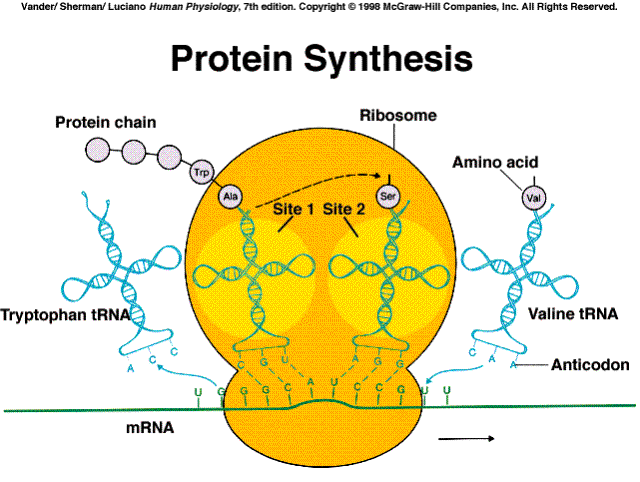
Structure of Ribosome - Biology Wise Diameter of Ribosome is 20nm. Their composition can be divided into two parts - 2/3 part of r-RNA (ribosomal RNA) and 1/3 part RNP (Ribosomal protein or Ribonuclep protein). Polypeptide chain is fabricated by translating mRNA (messenger RNA) with the aid amino acids that tRNA (transfer RNA) delivers.
Ribosome diagram with labels
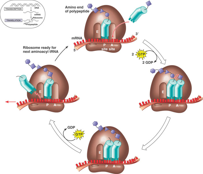
Active Ribosome Profiling with RiboLace - PubMed Ribosome profiling, or Ribo-seq, is based on large-scale sequencing of RNA fragments protected from nuclease digestion by ribosomes. Thanks to its unique ability to provide positional information about ribosomes flowing along transcripts, this method can be used to shed light on mechanistic aspects … Active Ribosome Profiling with RiboLace Solved In the following diagram of a ribosome, assign the - Chegg In the following diagram of a ribosome, assign the correct labels. Who are the experts? Experts are tested by Chegg as specialists in their subject area. We review their content and use your feedback to keep the quality high. Transcribed image text: In the following diagram of a ribosome, assign the correct labels. Ribosome - protein factory - definition, function, structure and biology The protein translation by a ribosome consists of three stages: (1) Initiation, (2) Elongation, and (3) Termination. Initiation - the ribosome assembles around the target mRNA. A small ribosome subunit links onto the "start-end" of an mRNA strand. "Initiator tRNA" also enters the small subunit and binds to the start codon (most commonly, AUG).
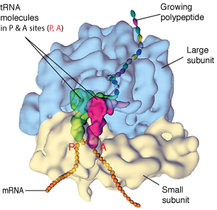
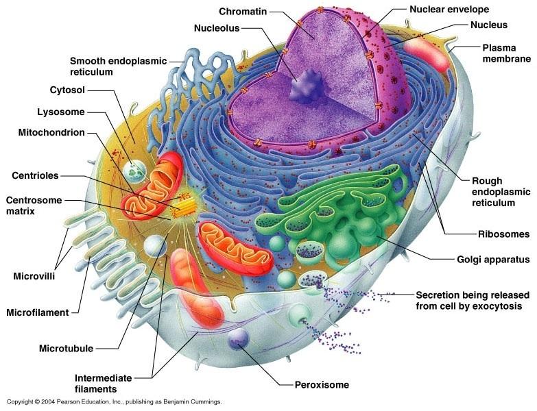
Ribosome diagram with labels. Animal Cell Diagram | Science Trends The diagram, like the one above, will include labels of the major parts of an animal cell including the cell membrane, nucleus, ribosomes, mitochondria, vesicles, and cytosol. The cells of animals are the basic structural units for the wide variety of life we see in the animal kingdom. Animal Cells: Labelled Diagram, Definitions, and Structure Ribosomes Ribosomes create proteins. They can float freely in the cytoplasm or can be attached to the nuclear envelope. They create proteins by assembling amino acids into polypeptides. As the ribosomes build an amino acid chain, the chain is pushed into the endoplasmic reticulum. What Are Ribosomes? - Definition, Structure and its Functions Ribosomes are located inside the cytosol found in the plant cell and animal cells. The ribosome structure includes the following: It is located in two areas of cytoplasm. Scattered in the cytoplasm. Prokaryotes have 70S ribosomes while eukaryotes have 80S ribosomes. Around 62% of ribosomes are comprised of RNA, while the rest is proteins. Bio 1113 - Unit 11 - Gene Expression Flashcards - Quizlet In the following diagram of a ribosome, assign the correct labels: Label 1: a tRNA attached to a polypeptide is found in this area of the ribosome Label 2: a tRNA attached to a single amino acid enters here Label 3: a tRNA that is not attached to anything exits here Label 4: a tRNA molecule Label 5: growing polypeptide Label 6: mRNA being ...
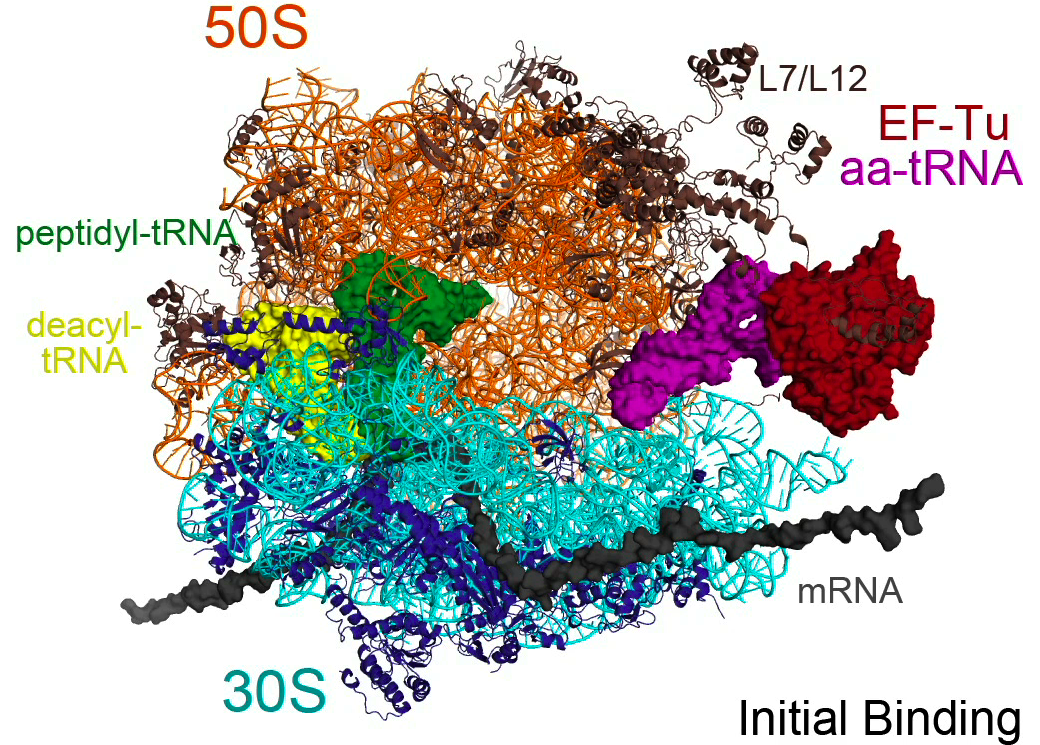
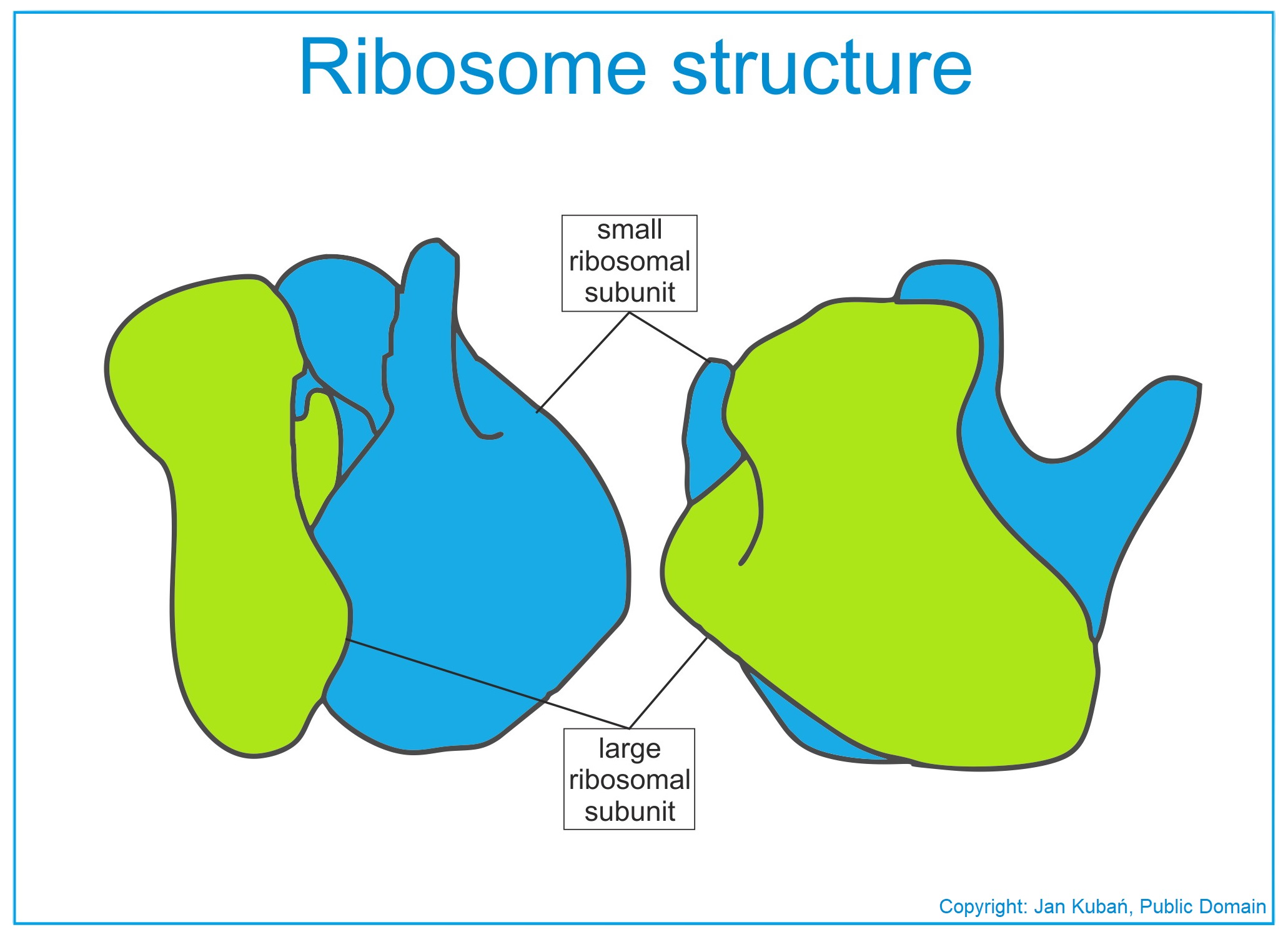
Ribosomes Stock Illustrations - 656 Ribosomes Stock ... - Dreamstime Download 656 Ribosomes Stock Illustrations, Vectors & Clipart for FREE or amazingly low rates! New users enjoy 60% OFF. 190,135,943 stock photos online. ... Anatomical and medical labeled scheme. Explained closeup diagram. Ribosomes vector illustration. Anatomical and medical labeled. Structure of Ribosome (With Diagram) - Biology Discussion A bacterial ribosome is about 250 nm in diameter and consists of two subunits, one large and one small. Both subunits consist of one or more molecules of rRNA and an array of ribosomal proteins. ADVERTISEMENTS: Association of two subunits is called mono-some. The structure of prokaryotic ribosome is given in the figure 8.2 B. Ribosome - Wikipedia The ribosomal proteins and rRNAs are arranged into two distinct ribosomal pieces of different sizes, known generally as the large and small subunit of the ribosome. Ribosomes consist of two subunits that fit together (Figure 2) and work as one to translate the mRNA into a polypeptide chain during protein synthesis (Figure 1). A Labelled Diagram Of Mitochondria with Detailed Explanation The ribosomes found within the mitochondria are called the mitochondrial ribosome or mitoribosome. It is a protein complex which functions by translating mitochondrial mRNAs encoded in mtDNA. Also Read: Ribosomes Inner membrane The inner mitochondrial membrane holds proteins and functions by permitting the entry of only the selected molecules.
Ribosomes Vector Illustration. Anatomical and Medical Labeled Scheme ... Ribosomes vector illustration. Anatomical and medical labeled scheme. Explained closeup diagram.. Illustration about amino, educational, biogenesis, labeled, golgi, body - 122097933 Labeled Plant Cell With Diagrams | Science Trends The ribosomes are created in the nucleolus of the cell. Ribosomes are made out of two smaller subunits, a large ribosomes subunit and a small ribosomal subunits. The transfer RNA or tRNA encodes the correct series of genetic instructions into the mRNA or messenger RNA, which is what ensures that the right proteins are created. Ribosome and protein synthesis, diagram - Stock Image - C029/3020 Ribosome and protein synthesis, diagram. C029/3020. Rights Managed. 50.0 MB (926.7 KB compressed) 4826 x 3621 pixels. 40.9 x 30.7 cm ⏐ 16.1 x 12.1 in (300dpi) This image is not available for purchase in your country. Please contact your Account Manager if you have any query. Request Price Add To Basket. Ribosomes- Definition, Structure, Functions and Diagram Ribosomes Definition The ribosome word is derived - 'ribo' from ribonucleic acid and 'somes' from the Greek word 'soma' which means 'body'. Ribosomes are tiny spheroidal dense particles (of 150 to 200 A0 diameters) that are primarily found in most prokaryotic and eukaryotic. They are sites of protein synthesis.
Chimera Animations
Blood Histology Slides with Description and Labeled Diagram Again, you will find the Golgi body, mitochondria, ribosome, rough endoplasmic reticulum in the cytoplasm of the eosinophil. The granules of the eosinophils may vary in different animal species. You will find numerous uniform, spherical granules in the eosinophil of sheep, goats, cows, and pigs. ... All the blood cell histology labeled diagrams ...

The Ribosome: Structure, Function and Location - Video & Lesson Transcript | Study.com
What is the name of the structure labeled Fin the diagram below? A ... Find an answer to your question What is the name of the structure labeled Fin the diagram below? A. Ribosome B. Cytoplasm C. Mitochondrion D. Nucleus brooke8378 brooke8378 02/02/2021 Biology ... owing questions on the basis of the given diagrams . ( i ) Sort out root , stem and leaf from the above . ( iv )
About Ribosomes class 9 science Biology Notes
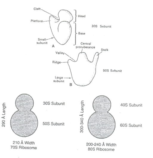
Cell Organelles- Definition, Structure, Functions, Diagram In the case of prokaryotic cells, the ribosomes are of the 70S with the larger subunit of 50S and the smaller one of 30S. Eukaryotic cells have 80S ribosomes with 60S larger subunit and 40S smaller subunit. Ribosomes are short-lived as after the protein synthesis, the subunits split up and can be either reused or remain broken up.
Translation Elongation
Ribosomes: Structure, Composition, and Assembly (With Diagram) Ribosomes in the cytoplasm of eukaryotic cells have a sedimentation coefficient of about 80 S (MW about 4.5 x 10 6) and are composed of 40 S and 60 S subunits. In prokaryotic cells, ribosomes are typically about 70 S (MW about 2.7 x 10 6) and are formed from 30 S and 50 S subunits.
What is a good analogy for a ribosome? - Quora
Bacteria in Microbiology - shapes, structure and diagram Bacterial spores. Bacterial endospores layers. Bacteria cells are the smallest living cells that are known; even though viruses are smaller than bacteria, viruses are not living cells. There are different types of bacteria with various sizes, shapes, and structures. The bacteria shapes, structure, and labeled diagrams are discussed below.
PROTEIN SYNTHESIS BASICS
Animal Cell Diagram with Label and Explanation: Cell ... - Collegedunia Animal cell is a typical Eukaryotic cell enclosed by a plasma membrane containing nucleus and organelles which lack cell walls, unlike all other Eukaryotic cells. The typical cell ranges in size between 1-100 micrometers. The lack of cell walls enabled the animal cells to develop a greater diversity of cell types.
Cells - Ms. Anderson's Biology Class
protein synthesis diagram labeled - TheFitnessManual Switch RNAs (tRNAs) deliver amino acids to the ribosome. - "protein synthesis diagram labeled" tRNAs are additionally RNA polymers. They're typically between 75 and 90 RNA nucleotides lengthy. However in contrast to mRNAs, that are linear, hydrogen bonding between nucleotides inside a tRNA causes it to fold up.

Ribosome Diagram Labeled - Data Diagram Medis
Ribosome - protein factory - definition, function, structure and biology The protein translation by a ribosome consists of three stages: (1) Initiation, (2) Elongation, and (3) Termination. Initiation - the ribosome assembles around the target mRNA. A small ribosome subunit links onto the "start-end" of an mRNA strand. "Initiator tRNA" also enters the small subunit and binds to the start codon (most commonly, AUG).
SOLVED:You already know that ribosomes are abunda…
Solved In the following diagram of a ribosome, assign the - Chegg In the following diagram of a ribosome, assign the correct labels. Who are the experts? Experts are tested by Chegg as specialists in their subject area. We review their content and use your feedback to keep the quality high. Transcribed image text: In the following diagram of a ribosome, assign the correct labels.
The Cell
Active Ribosome Profiling with RiboLace - PubMed Ribosome profiling, or Ribo-seq, is based on large-scale sequencing of RNA fragments protected from nuclease digestion by ribosomes. Thanks to its unique ability to provide positional information about ribosomes flowing along transcripts, this method can be used to shed light on mechanistic aspects … Active Ribosome Profiling with RiboLace
Ribosomes Stock Photos, Images, & Pictures | Shutterstock

ribosome structure | Plant cell, Cell wall, Prokaryotes

Period 2, Keener and Risser, History of Earth Timeline | Timetoast timelines

escor_ex "The world is yours": Structure-of-a-plant-cell
Transcription and Translation labelling - Labelled diagram
The 59 keys for understanding the beginning of life
Print Chapter 2 cells flashcards | Easy Notecards
The Structure Of A Ribosome Stock Vector Illustration 304860134 : Shutterstock
Post a Comment for "42 ribosome diagram with labels"