41 the human heart and its labels
Heart histology: Cells and layers - Kenhub The heart is composed of left and right atria and ventricles. The atria are superior to (and less muscular than) the ventricles. The right and left atria are receptacles for blood coming from the body and lungs, respectively. On the other hand, the right and left ventricles eject blood from the heart to the lungs and body, respectively. How the Heart Works: How Blood Flows, Parts of the Heart, and More - WebMD Right side of the heart. Blood enters the heart through two large veins, the inferior and superior vena cava, emptying oxygen-poor blood from the body into the right atrium. As the atrium ...
Anatomical Planes of Body - The Human Memory The X-axis is going from left to. right, Z-axis from front to back, and Y-axis from up to down. In anatomical. terminology, three references plane are considered standard planes; these. planes differentiate the body anterior and posterior, ventral and dorsal, dexter, and sinister portions. Let me tell you about these standard planes in detail.

The human heart and its labels
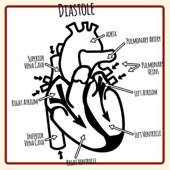
Heart - Life processes - Class Notes The sequence of events which take place during the completion of one heart beat is called cardiac cycle. It involves repeated rhythmic contraction and relaxation of heart muscles. Relaxation is called diastole. (1) Oxygen rich blood from the lungs enter the left atrium. The left atrium relaxes when it is collecting the blood. What Are the Four Main Functions of the Heart? - MedicineNet The heart is a muscular organ situated in the chest just behind and slightly toward the left of the breastbone. It roughly measures the size of a closed fist. The heart works all the time, pumping blood through the network of blood vessels called the arteries and veins. The heart and its blood vessels are known as the cardiovascular system. The Human Body: Anatomy, facts & functions | Live Science Each day, the kidneys process about 200 quarts (50 gallons) of blood to filter out about 2 quarts of waste and water. Adults excrete about a quarter and a half (1.42 liters) of urine each day. The ...
The human heart and its labels. Heart - Wikipedia The wall of the heart is made up of three layers: epicardium, myocardium, and endocardium. [7] The heart pumps blood with a rhythm determined by a group of pacemaker cells in the sinoatrial node. These generate a current that causes the heart to contract, traveling through the atrioventricular node and along the conduction system of the heart. 28 Amazing Facts About Your Heart - Cleveland Clinic These fascinating facts about your ticker may surprise you. You may not think much about your heart as it pumps away faithfully. But at Cleveland Clinic, we think about hearts a lot. We're proud ... Know the Structures and Functions about Your Heart The human heart is just roughly about the size of a fist. Your heart weighs about 10 - 12 ounces (or 280 - 340 grams) if you are a man, and 8 -10 ounces (or 230 - 280 grams) if you are a woman. In an adult, the heart beats at an average of 60-80 times per minute. The newborn's heart beats faster than an adult heart, at about 70-190 beats/minute. Double Circulation of Blood: Definition, Diagram - Embibe Ans: Double circulation is necessary for human beings because of the following advantages:-1. It checks the mixing of deoxygenated and oxygenated blood. 2. Oxygenated blood carries more oxygen which is supplied to all parts of the body. 3. Deoxygenated blood carries more carbon dioxide for its removal from the body. Q2.
Circulatory system - Wikipedia The circulatory system includes the heart, blood vessels, and blood. The cardiovascular system in all vertebrates, consists of the heart and blood vessels. The circulatory system is further divided into two major circuits - a pulmonary circulation, and a systemic circulation. The pulmonary circulation is a circuit loop from the right heart taking deoxygenated blood to the lungs where it is ... Heart: illustrated anatomy - e-Anatomy - IMAIOS 1 - RCA proximal 1. Basal anterior 10 - Second diagonal 10. Mid inferior 10a - Second diagonal a 11 - Proximal circumflex 11. Mid inferolateral 12 - Intermediate/anterolateral 12. Mid anterolateral 12a - Obtuse marginal a 12b - Obtuse marginal b 13 - Distal circumflex 13. Apical anterior 14 - Left posterolateral 14. Apical septal Heart Labeling Quiz: How Much You Know About Heart Labeling? Here is a Heart labeling quiz for you. The human heart is a vital organ for every human. The more healthy your heart is, the longer the chances you have of surviving, so you better take care of it. Take the following quiz to know how much you know about your heart. Questions and Answers 1. What is #1? 2. What is #2? 3. What is #3? 4. What is #4? Parts of the Heart & Blood Flow | Diagram & Overview - Study.com The four parts of the heart are the right atrium, right ventricle, left atrium, and left ventricle. The atrium receives blood from the body, the right ventricle pumps blood to the lungs, the left...
The Heart's Electrical System: Anatomy and Function The cardiac electrical signal controls the heartbeat in two ways. First, since each electrical impulse generates one heartbeat, the number of electrical impulses determines the heart rate. In normal sinus rhythm, that rate will be between 60 and 100 beats per minute. The sinus node signal also controls electrical conduction of the heart's steps ... Why the world of LGBTQ health doesn't fit under a single label According to the U.S. Office of Disease Prevention and Health Promotion, discrimination against LGBTQ people has been associated with high rates of psychiatric conditions, substance abuse and suicide. And a 2020 scientific statement from the American Heart Association, which Poteat and Streed helped write, said "there is growing evidence that ... 71 Interesting Facts About Human Heart - The Fact File There are four chambers (each chamber holds about 70 ml of blood) in the human heart. The total volume of the heart is therefore approximately 4 times that value, or 280 ml. There is upper right and left atria and there is lower right and left ventricle. Each of these chambers has a one-way valve which is positioned at its exit. Anatomy of the heart and coronary arteries (coronary CT) - IMAIOS Anatomical parts 1. Basal anterior 10. Mid inferior 11. Mid inferolateral 12. Mid anterolateral 13. Apical anterior 14. Apical septal 15. Apical inferior 16. Apical lateral 17. Apex 2. Basal anteroseptal 3. Basal inferoseptal 4. Basal inferior 5. Basal inferolateral 6. Basal anterolateral 7. Mid anterior 8. Mid anteroseptal 9. Mid inferoseptal
Two-Chambered Heart: Overview & Anatomy - Study.com The human heart is composed of four chambers: two ventricles and two atria. Deoxygenated blood enters the heart in the right atrium. From here, a valve opens, and blood enters the right ventricle ...

Kenya Forensics Online Resource: CARDIAC MUSCLE TISSUE
Layers of the heart: Epicardium, myocardium, endocardium - Kenhub Histologically, the heart is made of three layers of tissue: epicardium, myocardium, and endocardium. Contents Epicardium Myocardium Endocardium Clinical relations: Endocarditis Sources + Show all Epicardium Visceral layer of pericardium Lamina visceralis pericardii 1/2 Synonyms: Epicardium The epicardium is the outermost layer of the heart.
CLASS NOTES: Basic Heart Anatomy (Vital Signs: Understanding What the ...
The Human Heart - Anatomy & Passage Of Blood - TeachPE.com Anatomy of the heart The heart consists of four chambers and is divided into left and right by a wall of muscle called the septum. By convention the heart is often illustrated with the left side on the right as you look at it. In other words, imagine you are looking at someones heart; your right is their left! Atria & Ventricles
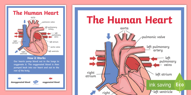
Cursive The Human Heart Display Poster (teacher made)
How the Heart Works - The Heart | NHLBI, NIH It is made up of multiple layers of tissue. Your heart is at the center of your circulatory system. This system is a network of blood vessels, such as arteries, veins, and capillaries, that carries blood to and from all areas of your body. Your blood carries the oxygen and nutrients that your organs need to work properly.
File:Diagram of the human heart.svg - Wikimedia Commons
Human heart: Anatomy, function & facts | Live Science The human heart has four chambers: two upper chambers (the atria) and two lower ones (the ventricles), according to the National Institutes of Health. The right atrium and right ventricle together...

Circulatory System
Quiz: Do You Know About The Anatomy Of The Human Heart? None of the above 4. Two large veins that drain blood from the upper body and the lower body and empty it into the right atrium of the heart are known as the: 5. Two of the four chambers with powerful muscular contractions that force blood to flow through arteries to all parts of the body: 6.
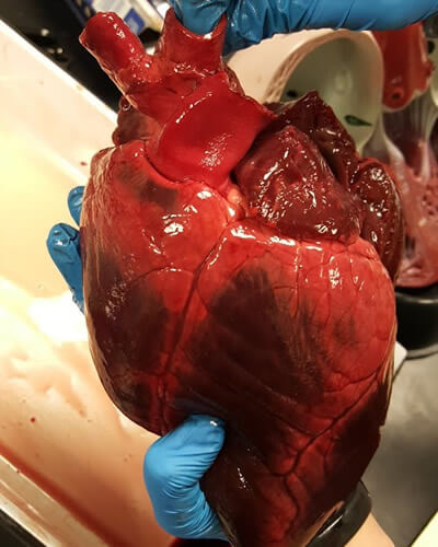
Deer Heart Dissection
How the Heart Works: Diagram, Anatomy, Blood Flow - MedicineNet The heart is located under the rib cage -- 2/3 of it is to the left of your breastbone (sternum) -- and between your lungs and above the diaphragm. The heart is about the size of a closed fist, weighs about 10.5 ounces, and is somewhat cone-shaped. It is covered by a sack termed the pericardium or pericardial sack.
.svg/399px-Diagram_of_the_human_heart_(no_text).svg.png)
File:Diagram of the human heart (no text).svg - Wikimedia Commons
The Human Heart Online Quiz | Science for Kids | 10 Questions 4. The heart of all mammals is divided into four chambers. Fish and reptiles have a 2-chambered heart. Tadpoles have a 2-chambered heart, but develop a 3rd chamber when they become adults. No animal has a 5-chambered heart. Some of the heart's chambers are located at the top and some at the bottom.
33 Human Heart To Label
Diagram of Human Heart and Blood Circulation in It A heart diagram labeled will provide plenty of information about the structure of your heart, including the wall of your heart. The wall of the heart has three different layers, such as the Myocardium, the Epicardium, and the Endocardium. Here's more about these three layers. Epicardium

KS2 Heart Diagram QR Labelling Activity - Science - Twinkl
Heart Drawing With Label : Human Heart Diagram Images Stock Photos ... Click here to get an answer to your question ️ draw a diagram of the human heart and label its parts. Cell structure and functions / animal cell vs plant cell / parts of cell / ch 8 science class 8 cbse . This is how to draw human heart diagram step by step.please like share and subscribe my youtube channel. How to draw human heart with ...

Human Heart Drawing With Labels / Anatomical Heart By ... - Didier Mireault Download this sketch of human heart anatomy with hand written labels vector illustration now. In this interactive, you can label parts of the human heart. Draw pulmonary aorta from center . Click here to get an answer to your question ️ draw a diagram of the human heart and label its parts. And search more of istock's library of .
Diagram Of The Human Heart Stock Images, Royalty-Free Images & Vectors ...
Total Artificial Heart - What Is Total Artificial Heart? | NHLBI, NIH A total artificial heart (TAH) is a pump that is placed in the chest to replace damaged heart ventricles and valves. (Ventricles pump blood to the lungs and other parts of the body.) Once the pump has been placed in the chest, a machine called a driver controls the pump outside the body. The pump and driver help blood flow to and from the heart ...

Longitudinal Section of a Kidney | ClipArt ETC
The Human Body: Anatomy, facts & functions | Live Science Each day, the kidneys process about 200 quarts (50 gallons) of blood to filter out about 2 quarts of waste and water. Adults excrete about a quarter and a half (1.42 liters) of urine each day. The ...

Anatomy of the Human Heart – Jansen’s Lessons
What Are the Four Main Functions of the Heart? - MedicineNet The heart is a muscular organ situated in the chest just behind and slightly toward the left of the breastbone. It roughly measures the size of a closed fist. The heart works all the time, pumping blood through the network of blood vessels called the arteries and veins. The heart and its blood vessels are known as the cardiovascular system.

34 Label The Human Heart - Labels For Your Ideas
Heart - Life processes - Class Notes The sequence of events which take place during the completion of one heart beat is called cardiac cycle. It involves repeated rhythmic contraction and relaxation of heart muscles. Relaxation is called diastole. (1) Oxygen rich blood from the lungs enter the left atrium. The left atrium relaxes when it is collecting the blood.

Human Heart Nomenclature - Cards | Human heart, February classroom ...


The Human Body Facts, Worksheets & Key Systems For Kids
Post a Comment for "41 the human heart and its labels"